Experiments with the blind spot and filling in
How to draw your blind spot and the blood vessels that obscure vision
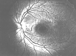
The Figure below is a photograph of the author's retina, seen from the front. From where they enter the eyeball, the blood vessels spread out to cover the surface of the retina in the way giant spiders might straddle each eye in a science fiction film. The blind spot is visible on the left side, as the body of the spider, from which the legs radiate out.
Under special conditions it is possible to make the blind spot and blood vessels visible[1]. In the method which is most effective, you take a strong penlight with a small, concentrated beam, and hold it very close to the side of the eye, so that its light creates a small bright spot on the white of the eye. This strong light traverses the cornea and creates a reddish glow on the inside of the eyeball. Since this glow is located to the side of the pupil, that is, at a spot inside the eyeball where usually no light comes from, the shadows of the retinal blood vessels that are cast by this glow are at an abnormal position and become noticeable, particularly when the flashlight is moved slightly[2].
What you see is quite similar to what is seen in Figure above. There is a large knot of vessels, corresponding to the blind spot, on the side away from the nose and slightly below the horizontal midline. Looking straight ahead however, that is, in central vision, there are not many vessels. This because at the very center of the visual field there is a region called the fovea where the photoreceptors are most concentrated and where vision is keenest. Presumably so that foveal vision can be as acute as possible, the web of blood vessels carefully skirts around this spot. The spot is called fovea ('depression', in latin) because under the microscope it appears as a slight dimple in the surface of the retina.
A simple way of showing that you actually see nothing in the region of the blind spot is the following. You close the left eye, for example, and look with the right eye at your left thumb, held at arm's length. (It is important to get the sides right, since the blind spot is situated asymetrically, on the nasal side of each retina, that is, the blind spot of the right eye is on the right of the line of sight and the blind spot of the left eye is on the left of the line of sight). Next to your left thumb you hold your right thumb, which you gradually move away from the left thumb, towards the right. At a separation between your thumbs of about 6 - 10 inches (corresponding to an angle at the eye of about 10 - 15 degrees), while you continue looking at the stationary left thumb, your right thumb will disappear from view (of course it will reappear if you look at it directly). By moving your thumb slightly, you can ascertain that the blind spot is not situated exactly on the horizontal midline, but about 2-3 degrees below it. You can also see that it is surprisingly large: its diameter is about 5-6 degrees horizontally and 7-8 degrees vertically[3]: about the side of 12 full moons[4] set next to each other. This means that the blind spot can be made to devour a lemon or small orange held at arm's length.
Given the size of the blind spot, it is surprising that it such great observers as Aristotle, Galen, and Al Hazen did not discuss it, and that it only became known after Mariotte (1668) caused a sensation with it at the Royal Society of London, and after King Charles II supposedly amused himself by visually decapitating his courtisans[5].
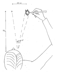
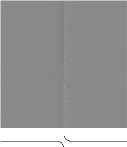
Helmholtz[6] gives an excellent method for drawing your own blind spot. You close one eye (say the left) and you look at a fixation point marked on a piece of paper. You localize the region of the blind spot to the right (when looking with the right eye) of the fixated point, and with a pencil, you blacken the paper over all the area for which the tip of the pencil remains invisible. As soon as the tip of the pencil comes out of the blind spot, you stop marking. By proceding in this way, and keeping the eye completely still (this is difficult!), it is possible to draw the vertically extended patch corresponding to the blind spot, and even to begin to draw the stumps of the two blood vessels that leave the spot at the top and at the bottom. The technique is illustrated below, on the left.
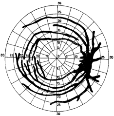
The Figure on the right (from Weekers, 1945) is the result
of performing just the same kind of mapping of the visual field, but with more
precision, and in conditions of weak illumination so we can descern not only
regions that are completely blind, like the blind spot, but also regions that
are less sensitive to light because they are obscured by blood vessels. The
figure is surprising, because it demonstrates both how vast the regions are
that are obscured by the web of retinal blood vessels, and also the extent to
which we are ignorant of these vessels. Why is it that we do not ordinarily
notice this vast web of vessels casting shadows all over the visual field? Why
don't we see the world as though we were peering out from behind the blood
vessels? Why dont we see the blind spot like the body of a giant spider, from
which the legs radiate out? The absence of blood vessels at the fovea explains
why vision is not deteriorated in the center of the visual field, but why does
one not see a large empty blob corresponding to the blind spot, off to the side
of each eye? Why does one need to resort to artificial methods like Helmholtz's
method described above, in order to become aware of the large gaps in our
visual field
White eye vs. red eye in flash photography
Another interesting
thing about the blind spot is that you can actually see it from outside
someone's eye. To understand this, consider the well-known effect of 'red-eyeÓ
when you take a flash photograph of someone. The reason people's eyes look red is
that the light from the flash travels into the eye, gets reflected by the
retina, and comes out again along the same path that it went in. When the
camera lens is positioned very close to the flash, as is usually the case in
amateur photography, this reflection is recorded on the film. Now, since the
retina is covered with blood vessels and photoreceptors, it absorbs most of the
light, and only reddish light comes back out. But at the blind spot, nerve
fibers bunch together to get out of the eye. There are no photoreceptors, no
light absorption, and, so most of the light landing on the blind spot simply
gets reflected. This means that if you were to take a flash photograph of
someone who had placed their eyes with their blind spot exactly at the position
of the flash, the photograph should show 'white-eyeÓ instead of 'red-eyeÓ. And
indeed this is what happens, as can be seen in the picture below

Red eye and white
eye! My son and daughter have placed their eyes in such a manner that their
left eye's blind spot is positioned on the flash. For this reason a bright
white spot appears on their left pupil, whereas their right pupil shows the
usual, weaker, red-eye effect deriving from reflection of the flash off a
different portion of the retina. The chances of this occurring unintentionally
are small, which explains why one rarely sees 'white-eyeÓ in photographs.
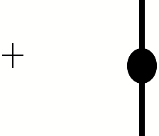
The disappearing lollipop trick
Close your left eye
and look at the cross on the left. Gradually move the book closer and closer to
your eye (be careful: make sure you have the LEFT eye closed and the right eye
open). Suddenly, at a distance of about
15 cm, the lollipop on the right will disappear, only the stick will
remain[7].

If you redo the
disappearing lolipop trick described above, but with a two-handled lolipop as
shown below, something curious happens. Now although the heart of the lolipop
disappears as before, the handles join up to make a single line that appears to
cross the blind spot.
Another interesting
test of the blind spot's intelligence is flowery wall-paper. If the flowers are
fairly small, so that they form an overall texture, you will find that you
don't have the impression that there is anything missing in the blind spot.
Admittedly it's hard to tell exactly what is present there, but you certainly don't have the impression that
there is an interruption of the surrounding flower-texture.
On the other hand if
the flowers are fairly big, so that only a single flower will fit into the
blind spot, then you definitely have the impression that there is a missing
flower.
A great debate has arisen in the vision scientist
community about experiments like this. Many vision scientists suppose that the
phenomena must be explained by an active filling-in mechanism that takes the
visual image and actually 'paints inÓ the information that is missing by
starting at the boundaries of the blind spot and spreading inwards[8].
Indeed vision
scientists have additional reasons for thinking that there should be such a
spreading activation mechanism. It seems that the perception of surface color
and surface brightness depend critically on the properties of the edges and
boundaries that delimit the colors.
A classic example is
the so-called Cornsweet-Craik-O'Brien illusion you see in the following Figure.
Cornsweet-Craik-O'Brian
illusion. Even though the left half looks darker, in fact the two halves of the
square are the same lightness except at the central boundary. The boundary is
constructed in a special way—it has a luminance profile as shown in the
graph under the figure: just as you reach the boundary coming from the left,
the lightness decreases slightly, then jumps up to a slightly higher value, and
then goes down to the same value that it was at the left of the Figure.
You have the
impression of seeing two surfaces of different lightness. But in fact they are
equally bright. You can convince yourself of this by putting a pencil in such a
way as to hide the boundary between the surfaces. The explanation is that the
boundary between the two surfaces has a special profile that induces the
effect.
Here are two more
examples showing that the perception of the color and lightness of surfaces
depends on the presence of abrupt changes in luminance. In these cases illusory
bright surfaces seem to be generated by nearby abutting lines.
These examples are
compatible with the existence of neural brightness-filling mechanisms that work
inwards from luminance discontinuities. The filling-in of the blind spot may be
another manifestation of such a mechanism.
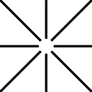
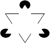
On the left, the
Ehrenstein illusion: The abutting lines create the effect of a
whiter-than-white disk floating above the background. On the right, the Kanizsa
triangle: The white triangle formed by the pacmen and the interrupted black lines
seems whiter than the white of the page, and seems to have a real boundary,
whereas in fact there is none.
Other scientists on
the other hand have a different interpretation of all these effects. Consider
the following set of dogs. Clearly the dog behind the wall is much longer than
his friends. Interestingly however you don't have the impression of seeing the whole length of the partially hidden dog.
You somehow know or sense that he is longer, but you don't have the
feeling of his belly being actually painted in. The brain seems to somehow assume that the rest of the dog is there, behind the
wall, without actually recreating it. This phenomenon is called 'amodal
completion', and seems to be a less visually 'present' form of filling in than
what we observe in the virtual contours and bright areas of the
Cornsweet-Craik-O'Brien, the Ehrenstein and the Kanizsa illusions.
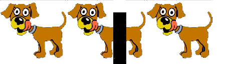
Amodal completion
of a dog. Even though the middle dog's tummy is hidden, one has the almost
palpable impression that it really is there.
Could it be that
what happens in the blind spot is more like amodal completion than true
active 'painting in' of missing information? The brain would have to do
less work that
way, since all that is necessary is that it should assume that there is the same kind of stuff in the
blind spot as nearby, and no active painting-in has to occur. This also makes
sense to the extent that we can't really see very accurately what is in
peripheral vision anyway.
The "homunculus" objection doesn't apply so much to
this type of interpretation of filling-in: Under this 'assuming it is there'
hypothesis, no internal 'picture' of the missing parts of the scene are created
for some internal homunculus to contemplate, since the information the observer
has about the missing parts of the scene is more a form of knowledge than a
real visual 'presentation'. But while this seems to square quite well with the
subjective impression you get in the case of amodal completion, there are cases
like the virtual contours or the brightness filling-in, where the feeling is
somehow more 'real' or 'visual'.
Despite homuncular
doubts, the idea of filling-in seems eminently reasonable, and can be found in
virtually every textbook on vision. Clearly filling-in, be it the active type
or the amodal type, exists
in the sense shown in the illustrations above.